Research software for adult spinal deformity

See the load above the fusion.

Zyra turns the standing radiographs you already take into a patient-specific model of the spine and its muscles, then estimates the compression and shear at the first mobile level above the construct.

  • In Standing AP and lateral films
  • Out Junctional compression and shear
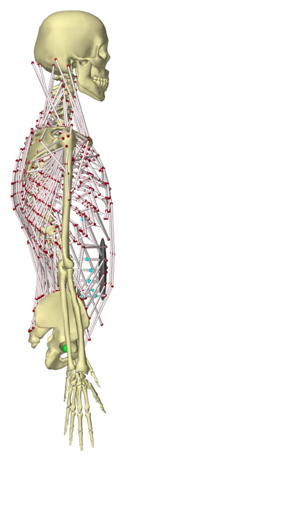
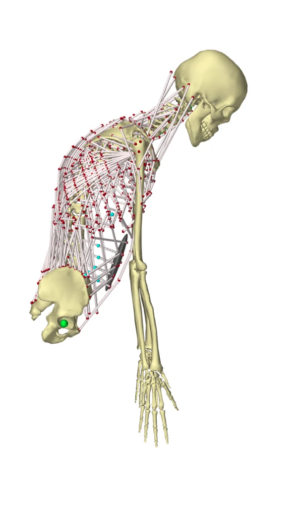
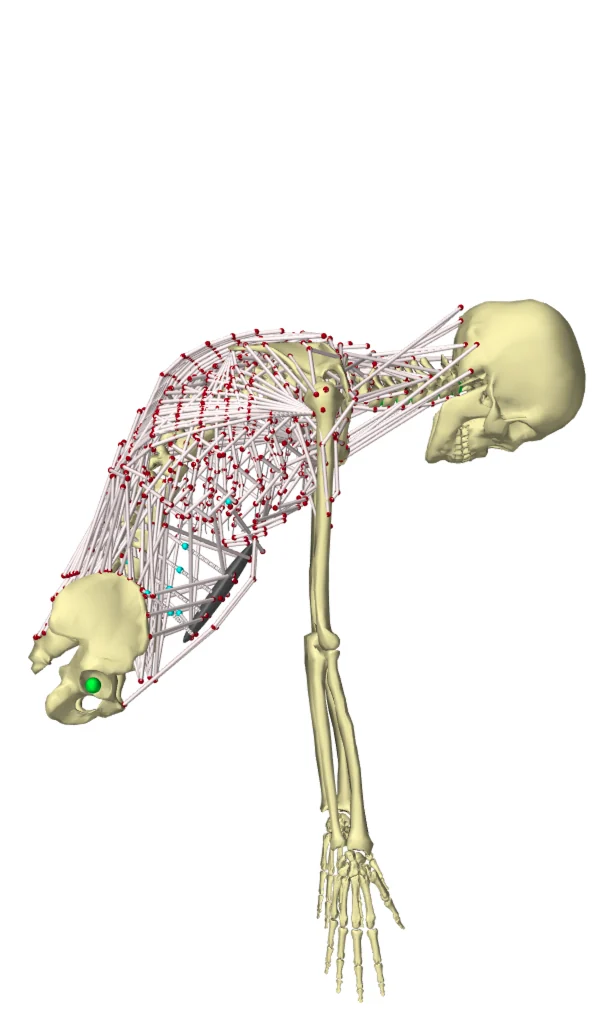
Illustration. As the trunk flexes, the muscles pull harder to hold it, and the first free level above the rod carries more of the load.

The problem

Alignment targets score the shape. They don't measure the load.

Proximal junctional failure is one of the most common reasons adults return to the operating room after deformity surgery. The frameworks used to plan these operations, from SRS-Schwab to GAP and age-adjusted targets, describe the shape of the corrected spine.

Two patients with the same alignment can still load the top of their construct very differently, because their muscles, body mass and posture differ. That load is what the junction actually has to carry, and today nobody sees it.

How it works

From two films to a load at every level.

Everything runs from imaging a deformity patient already has. No CT, no MRI, no motion lab.

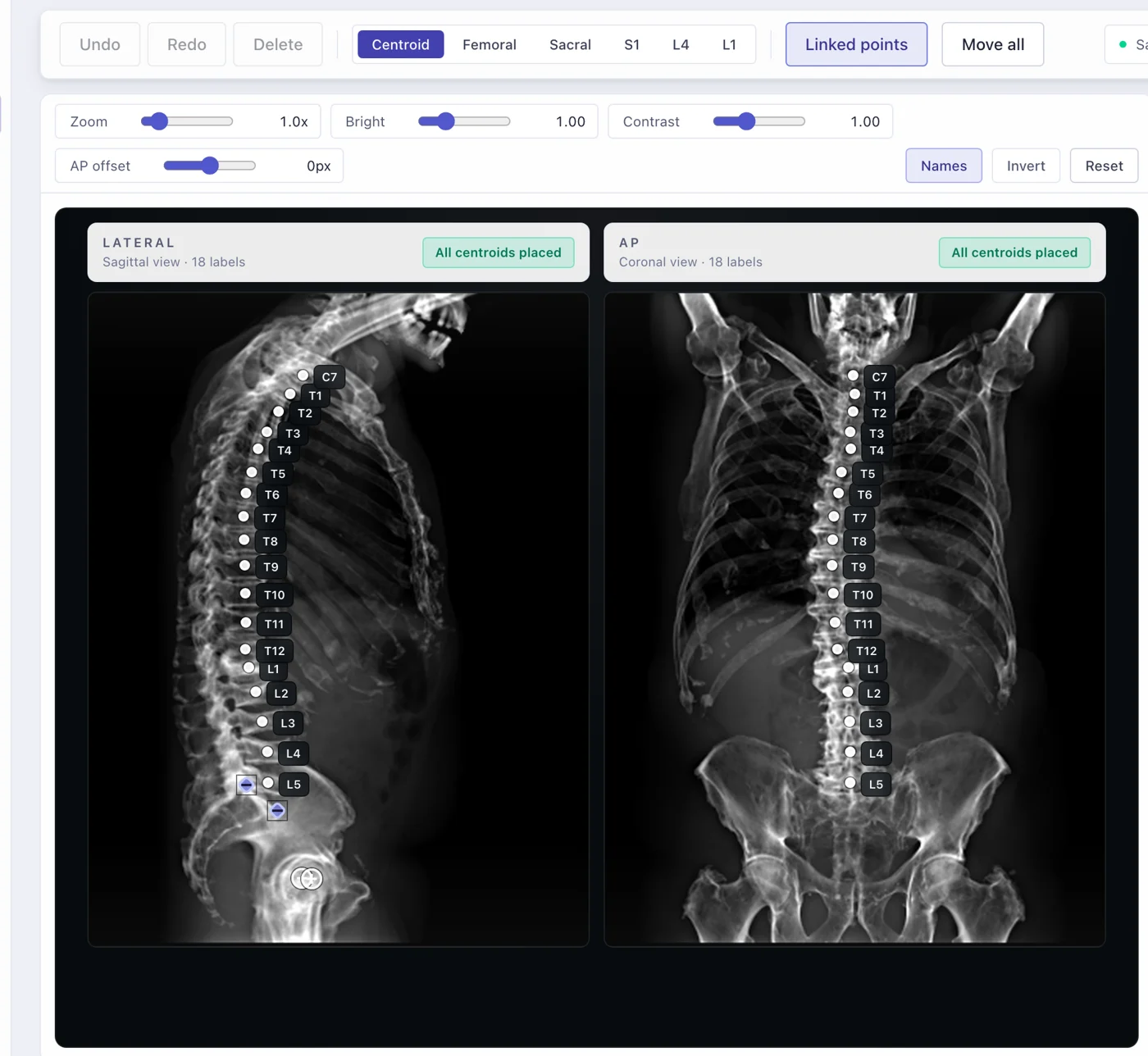
  1. Zyra landmarking view: lateral and frontal radiographs with vertebral centroids labelled from C7 to L5
    01

    Import the films, check the landmarks

    Drop in the standing AP and lateral radiographs as DICOM, JPEG or PNG; pixel size comes from the DICOM header or a quick calibration. A detector proposes vertebral centroids from T1 to L5 on both views, and you move anything that is off.

  2. 02

    Build the patient's model

    Landmarks from both views are reconstructed in 3D. A full thoracolumbar musculoskeletal model, with hundreds of muscle fascicles, is scaled to the patient and fitted to their spine, vertebra by vertebra.

  3. 03

    Estimate the junctional loads

    The model stands and bends through everyday tasks while the muscles do the work of holding each posture. Zyra reports compression and shear at the level above the fusion, before and after surgery, side by side.

Where it fits

Two moments in a deformity case.

First indication · in development

After surgery: who needs a closer look

Run the first standing film after surgery through the model. Junctional loads are estimated for that patient, so follow-up attention can go where the mechanics say it is needed.
Next

Before surgery: compare candidate plans

Choose a construct and an alignment goal, let the solver fit the corrected spine, and send each candidate through the same model. Compare the loads at the junction before you choose.

The workstation

One place for the film, the plan and the forces.

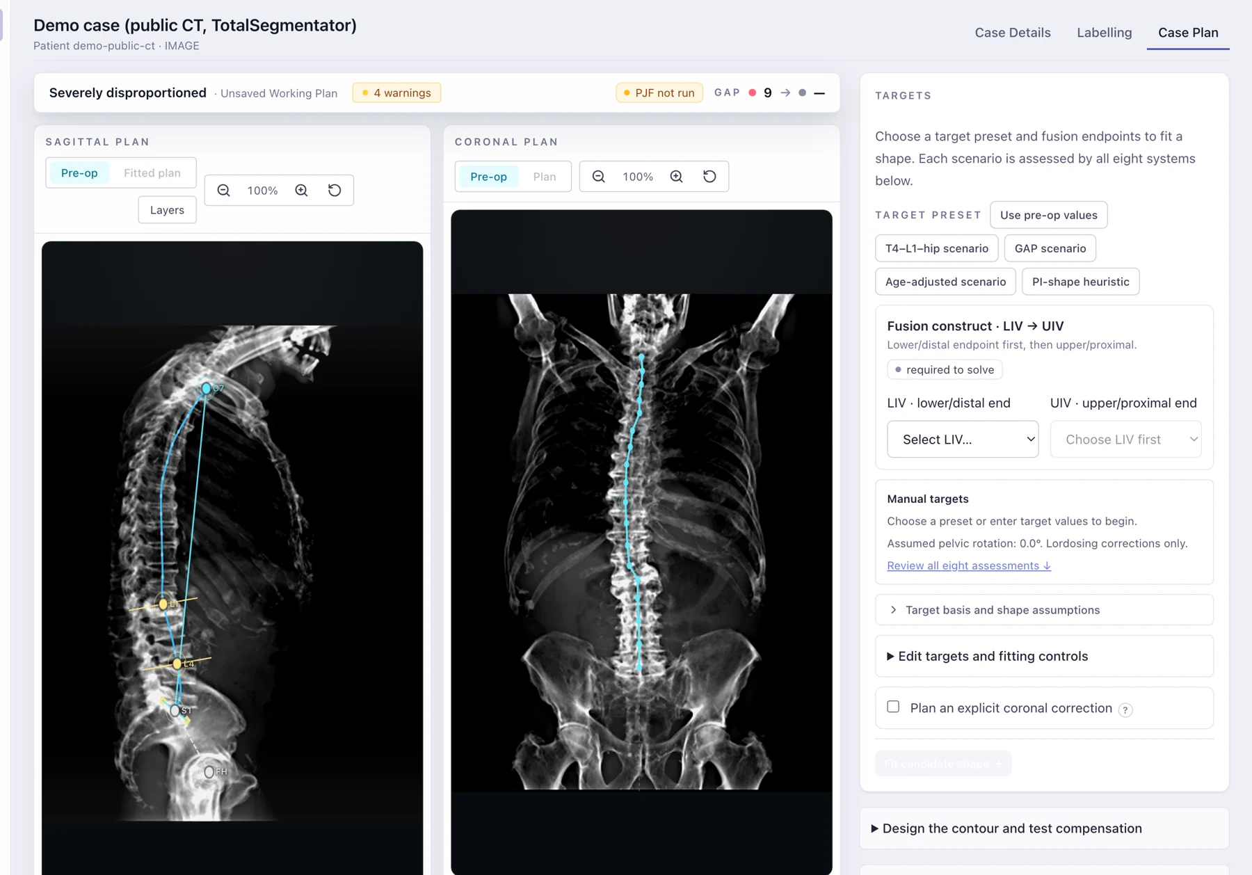
Zyra is a full planning workstation in the browser. The colours mean one thing each: cyan is measured from the film, orange is planned, and everything else stays out of the way of the radiograph.

Zyra plan workspace: sagittal and coronal plan views over the radiographs, with target presets and the fusion construct picker
Demo case. Radiographs were simulated from a public CT (TotalSegmentator, Wasserthal et al. 2023, CC BY 4.0). The CT was taken supine, so the alignment values are illustrative only.
  • Calibrated import

    DICOM, JPEG or PNG, with pixel size from the header or a draw-a-known-length tool.

  • Automatic landmarks

    T1 to L5 centroids proposed on both views, with every point reviewable.

  • Spinopelvic parameters

    PI, PT, SS, lumbar lordosis, global tilt, SVA and the GAP score, defined and documented.

  • Eight frameworks at once

    Every plan is assessed against eight established alignment systems, side by side.

  • Inverse plan solver

    Pick the fusion levels and a goal; the solver fits a corrected spine to match it.

  • Junctional force run

    Send the pre-op and planned spine to the biomechanics engine and read the loads back.

  • Printable case report

    Measurements, the plan and its alignment scores on one page for the case conference.

  • Built for hospital IT

    Single sign-on, organisation-scoped access, and an audit trail of every case read and write.

Science

Built on peer-reviewed work.

Each link in the chain from radiograph to load has been tested on its own. The outcome study, which asks whether these loads separate patients who later fail from those who don't, is under peer review.

  1. 2026
    Sensitivity of spinal load estimates to vertebral landmark error in biplanar radiograph–based musculoskeletal models

    Computer Methods in Biomechanics and Biomedical Engineering. How accurate the landmarks must be for the loads to hold.

  2. 2026
    Machine learning outperforms anthropometric scaling in predicting muscle parameters and spinal loading

    European Spine Journal. Patient-specific muscles without a CT or MRI of the trunk.

  3. 2025
    Subject-specific musculoskeletal modeling: the future of predicting and preventing proximal junctional failure in adult spinal deformity

    JOR Spine 8(4):e70142. The case for junctional loads, written with spine surgeons and biomechanists from several centres.

  4. 2025
    Effects of using generic vs. subject-specific muscle properties on spinal load prediction across different posture simulations

    Journal of Biomechanics 188:112741. Which muscle properties change the predicted load, and by how much.

About

From a thesis to a tool surgeons can use.

Zyra grew out of PhD research in biomedical engineering at the University of British Columbia on subject-specific musculoskeletal models of the spine in adult spinal deformity. The question behind it is simple: can a model built from routine imaging see the mechanical load that alignment rules miss?

Nima Ashjaee, founder. Biomedical engineer working where medical imaging, musculoskeletal modelling and machine learning meet. ashjaee.com

Research pilots

Working on adult deformity? Let's test it on your cases.

We are looking for spine surgery groups to take part in retrospective validation and research pilots. Tell us about your service and the data you hold, and we'll set up a call.